
FIGURE 2.
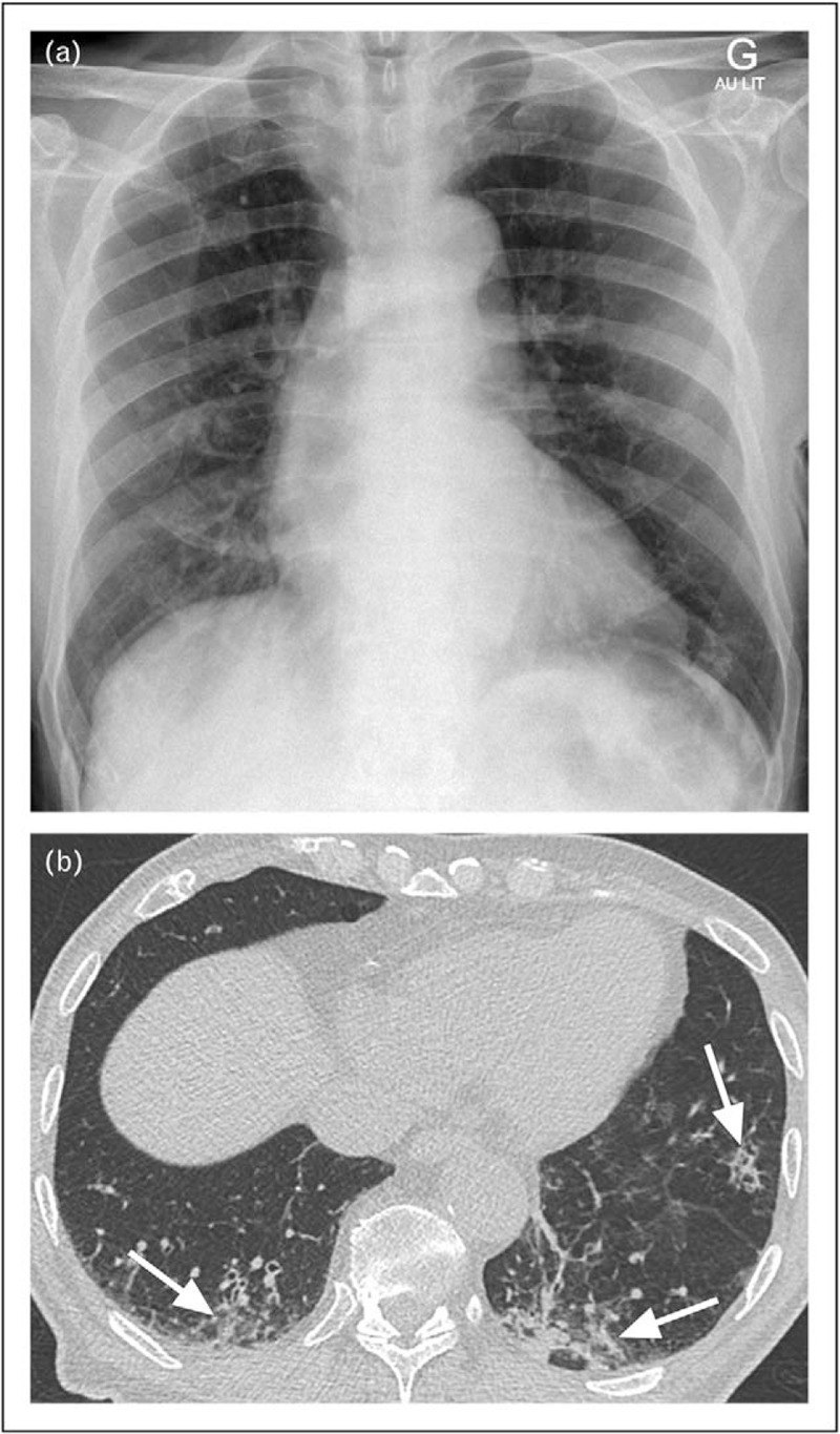
Images of 77-year-old man with suspicion of community-acquired pneumonia. a and b, frontal bedside chest radiograph (a) and unenhanced low-dose CT image of lung bases in lung window setting (b). No consolidation is obvious on the chest radiograph (a). The CT, however, shows faint infiltrates (arrows, b) affecting also nondependent lung regions. CT, computed tomography.