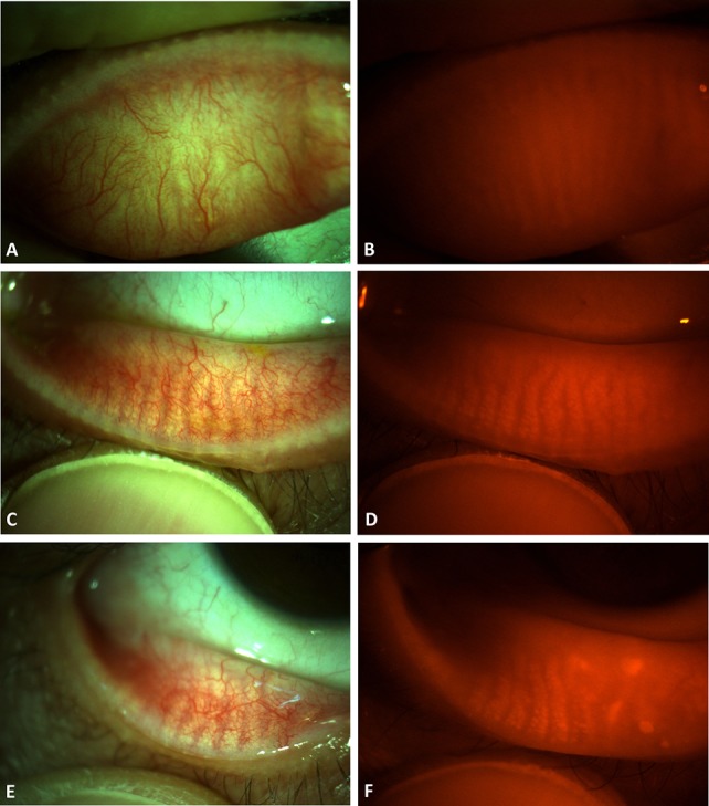
FIGURE 1.

Anterior segment color digital photographs of the eyelids with or without the red filter. In the upper (A and B) and lower (C and D) eyelids of the right eye of a 23-year-old female patient, the MGs can be visualized by simply inserting a red filter in front of the objective lens of the slit lamp (B and D). In the lower (E and F) eyelid of the right eye of a 53-year-old male patient, the MGs showed dropout in the nasal half.
