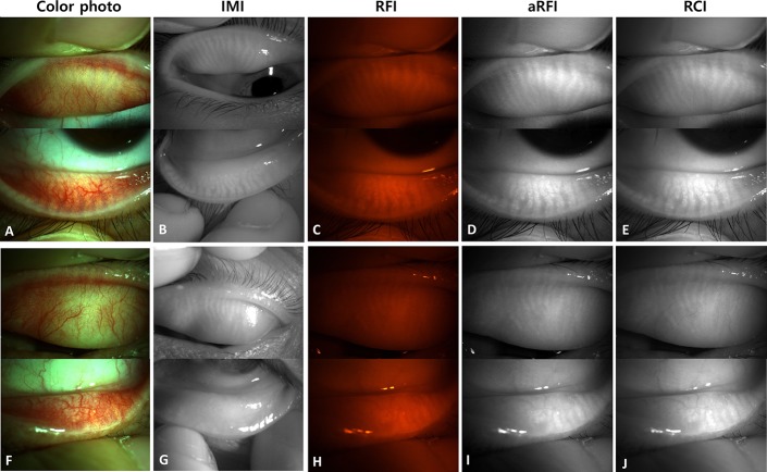
FIGURE 2.
Four sets of images of the tarsal conjunctiva and the underlying tarsus containing MGs. A–E, Images of the right eye of a 26-year-old female patient show intact MGs. F–J, Images of the left eye of a 56-year-old male patient show impaired MGs. A and F, Images of color photographs. B and G, Images of infrared meibography. C and H, Images acquired through a red filter. D and I, Brightness/contrast-adjusted images acquired through a red filter. E and J, Red-channel images of color photographs.

