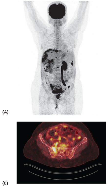
Figure 1.

Maximum intensity projection (A) and axial fused positron emission tomography/computed tomography (B) images of a 47-year-old patient with stage 1B serous ovarian carcinoma (patient no: 24) show widespread peritoneal involvement and mesenteric implants (SUVmax: 15.5), lymph nodes (SUVmax: 7.4), hypermetabolic lytic lesions in the sacrum and L3 vertebra (SUVmax: 16.5) suggestive of metastasis. The patient received chemotherapy, her serial positron emission tomography/ computed tomography images showed regression and CA-125 levels decreased progressively
