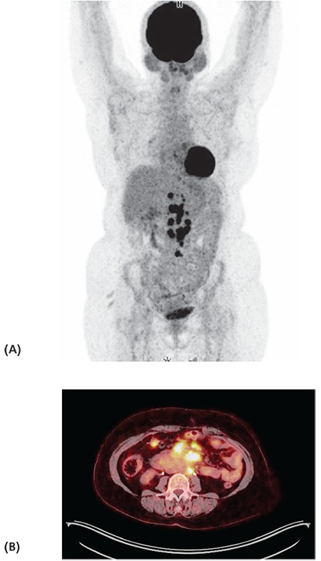
Figure 2.

Maximum intensity projection (A) and axial fused positron emission tomography/computed tomography (B) images of a 49 yearold patient with serous carcinoma (patient no: 26) show increased 18F-FDG uptake in para-aortic and celiac lymph nodes (SUVmax: 18.3) and mesenteric implants (4.2). Peritoneal biopsy confirmed malignancy in this patient
