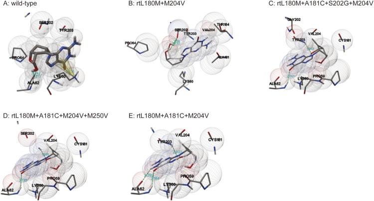
Figure 4.
Three dimensional structures of the entecavir triphosphate-binding domains of viral reverse transcriptase (RT). The effects of ETV-resistance mutations on the binding ability of HBV RT to ETV-TP were evaluated using a homology model constructed based on the crystal structure of HIV RT. The binding domains of a wild-type and four individual mutants are presented in the order of A, B, C, D, and E. Spheres represent HBV molecular surfaces. Green dot lines represent hydrogen bonds, and yellow net represents pi-cation interaction.

