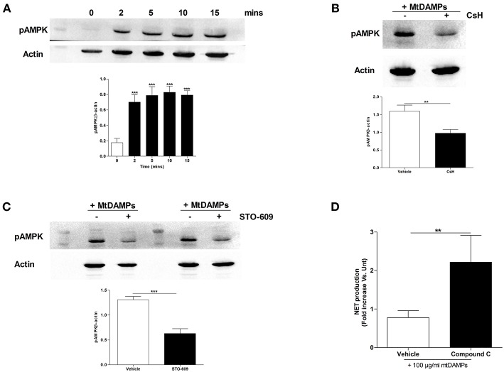
Figure 7.
Treatment of neutrophils with mtDAMPs results in phosphorylation of AMPK. (A) Whole cell lysates prepared from purified neutrophils stimulated for 2–15 min with 100 μg/ml mtDAMPs were screened for phosphorylated AMPK. Western blot in top panel is representative of 4 independent experiments. For densitometry analysis ***p < 0.0001 vs. 0 min. (B) AMPK phosphorylation in neutrophils treated for 1 h with the FPR-1 antagonist Cyclosporin H (CsH) or (C) or the CaMKK inhibitor STO-609 prior to a 5 min stimulation with 100 μg/ml mtDAMPs. Blots are representative of 5 (B) and 10 (C) independent experiments, with densitometric data depicted in the accompanying histogram. **p < 0.001, ***p < 0.0001 vs. vehicle. (D) Comparison of PMA-induced NET formation by mtDAMP stimulated neutrophils pre-treated with the AMPK inhibitor compound C or vehicle control (n = 10). **p < 0.01 vs. PMA treated.

