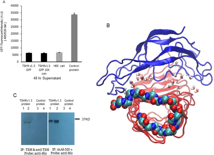
Figure 4.
TSH and TSHR antibody interaction with TSHRv1.3 protein. (A) Bar graph indicating quantitation of GFP fluorescent intensity for TSHRv1.3 protein in culture supernatants in unconcentrated and ×10 Amicon filter–concentrated supernatant from confluent cultures of stable HEK clones. The bar on the right is control protein GFP (1:100), which acted as the positive control. These data confirm the inability of the splice variant TSHRv1.3 protein to be secreted into the supernatant. (B) Molecular dynamic simulation (MD) of the TSHRv1.3 homology-modeled protein with TSH-β protein was performed using the CHARMM-GUI server for 200 ns. Shown here is a representative pose of the server-generated topology of the two complexed proteins. The β-pleated sheets and loops of α helices of TSH-β (blue) are seen to clasp the concave surface of the TSHRv1.3 ECD. Water molecules added to ensure neutrality are shown here as small stick structures within the complexed protein. The 21 amino acid intron 8 region of TSHRv1.3 is represented as continuous spheres of carbon (turquoise), nitrogen (blue), and oxygen (red). The MD simulation demonstrated that this truncated isoform of the receptor maintains the structural integrity to bind TSH with good stability. However, the affinity of binding was hard to predict in this modeling. (C) The binding of TSH and MS-1 to the purified protein was confirmed by immunoprecipitation (IP) with anti-TSH and anti-His antibodies as probes. The bound TSH-TSHRv1.3 complex formed by preincubation was precipitated with anti-TSH (10 μg per reaction) and the binding of MS-1 (10 μg per reaction) to TSHRv1.3 (10 and 50 μg protein per reaction, shown in lane 1 in the left-side blot and lane 2 in the right-side blot, respectively) was demonstrated with direct binding to purified protein followed by pull down of the complexed protein, using magnetic protein G beads. Normalized control protein lysate from vector-only transfected cells did not show any precipitation either with anti-TSH or MS-1 (lanes 3 and 4 in left and right blots, respectively). Therefore, this expressed receptor splice-variant protein has the ability to bind TSH and TSHR-stimulating antibody. mAb, monoclonal antibody.

