Figure 2.
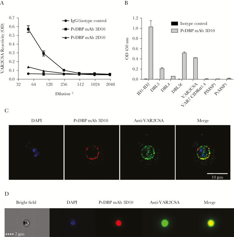
A Plasmodium vivax Duffy binding protein (PvDBP) mouse monoclonal antibody recognizes the Plasmodium falciparum surface antigen VAR2CSA. A, Titration of 2 PvDBP mouse monoclonal antibodies (mAbs; 3D10 and 2D10) and control immunoglobulin G1 (IgG1) against full-length VAR2CSA (FCR3) by enzyme-linked immunosorbent assay (ELISA). The concentration of the first dilution (1:50 on the x-axis) of each antibody was 8.6 μg/mL. Data are mean ODs (±SD). B, PvDBP mAb 3D10 and the isotype control (8.6 μg/mL for both) were tested against various recombinant Duffy binding–like (DBL) domains from VAR2CSA (ID1-ID2, DBL3X, DBL4ε, and DBL5ε), full-length VAR2CSA, IT4var07 CIDRα1.4, against P. falciparum MSP1 (PvMSP1), and against P. vivax MSP1 (PvMSP1) by ELISA, all coated at 0.5 μg/mL. C, red blood cells (RBCs) infected with mature P. falciparum strain CS2 parasites were fixed and costained with DAPI (blue), PvDBP mAb 3D10 (red), and a rabbit polyclonal antibody to VAR2CSA (green). Controls are shown in Supplementary Figure 2. D, Live cell images of a representative CS2-infected RBC (under bright field and with DAPI costaining) that shows costaining of PvDBP mAb 3D10 (red) and anti-VAR2CSA antibody (green).

