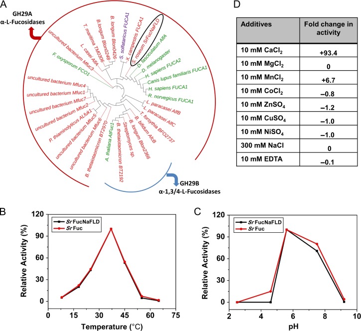
Fig. 3.
(A) Phylogenetic tree of SrFuc domain along with representative GH29-A and B alpha-l-fucosidase family members. Branch nodes and labels are designated in red for bacterial members, in green for eukaryotic members and in purple for the archaeal member. SrFucNaFLD is circled in black. (B) Enzymatic activity of SrFuc protein at different temperatures. Relative activity (%) values were calculated with respect to the highest value (enzyme activity at 37°C). (C) Enzymatic activity of SrFuc protein at different pH. Relative activity (%) values were calculated with respect to the highest value (enzyme activity at pH 5.6) (D) Enzymatic activity of SrFuc protein in the presence of metal ions, NaCl and EDTA. ND: not detected. Fold change in activity was calculated with the formula, (M–C)/C where M refers to activity in presence of metal ions and C refers to activity in a control tube lacking metal ions.

