
Figure 1. Iron prevents the cytotoxic effects of MO-OH-Nap treatment in MM cells.
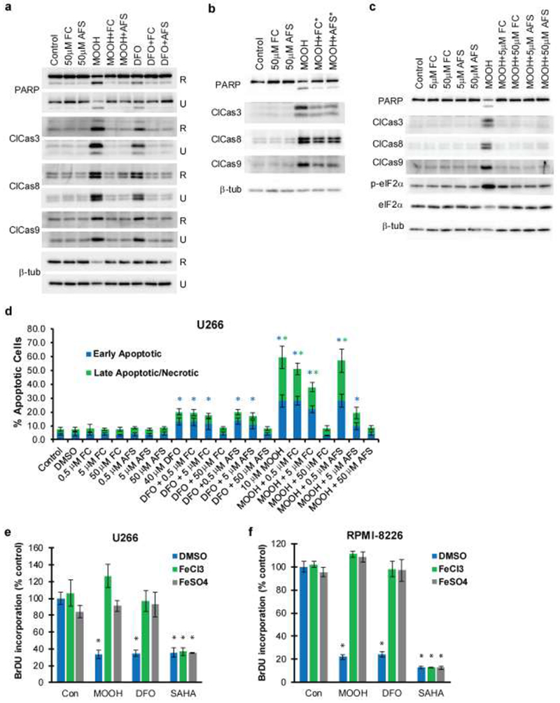
a) U266 (U) and RPMI-8226 (R) cells were treated for 48 hours with solvent control (DMSO), MO-OH-Nap (denoted as MOOH; 10 μM for U226 and 2.5 μM for RPMI), or 40 μM DFO in the presence or absence of 50 μM FC and 50μM AFS. Immunoblot analysis of cell lysates was performed using antibodies to detect PARP, cleaved caspases (3, 8, and 9) and β-tubulin (loading control). b) U266 cells were treated for 48 hours with solvent control or 10 μM MO-OH-Nap. 50 μM of AFS or FC were added 24 hours after the addition of MO-OH-Nap. PARP, cleaved caspases (3, 8, and 9) and β-tubulin were detected by immunoblot. c) U266 cells were treated with DMSO or 10 μM MO-OH-Nap in the presence or absence of either 5 μM or 50 μM FC or AFS for 48 hours. Protein levels of PARP, cleaved caspases (3, 8, and 9), p-eIF2a, eIF2a and β-tubulin were assessed by immunoblot. Immunoblots (a-c) are representative of three independent experiments. d) U226 cells were treated for 48 hours with FC, AFS, MO-OH-Nap (10 μM), and DFO (40 μM). Percentage of early apoptotic and late apoptotic/necrotic cells were determined via flow cytometry. Early apoptotic cells are defined as AnnexinV+,PI− while late apoptotic/necrotic cells are defined as AnnexinV+,PI+. Data are shown as mean ± S.D. (n=3) * denotes p<0.05 for comparison to the vehicle-only control sample. e) BrdU incorporation was measured in U266 and RPMI-8226 cells treated with MO-OH-Nap (2.5 μM for RPMI-8226 and 10 μM for U266), 40 μM DFO, or 1 μM SAHA in the presence of solvent control (DMSO) or 50 μM AFS/FC. Data are shown as mean (normalized to control) ± S.D. (n=3). *denoted p<0.05 for comparison to the vehicle-only control.