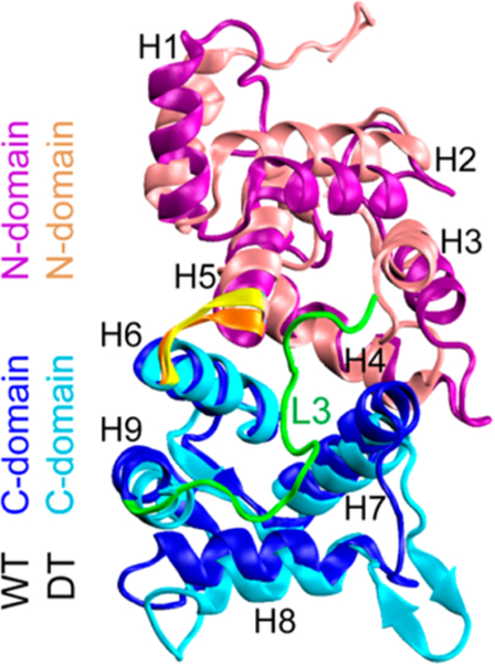
Figure 4.

Cartoon representations of WT NCS-1 and its DT variant superimposed on each other. The WT NCS-1 is more compact than the DT variant. The two structures were taken respectively from the MD trajectories of WT1 and DT1 at t = 500 ns. The N-domain and C-domain segments are shown respectively in purple and blue for WT species and in pink and cyan for DT NCS-1. The hinge loops are showed in orange and yellow for WT and DT protein. Loop L3 segment is shown in green for WT NCS-1.
