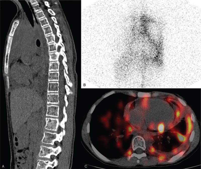
Figure 1.

99mTc-sulfur colloid (SC) lymphoscintigraphy and single photon emission computed tomography/computed tomography (SPECT/CT). (A) Sagittal CT showed multiple osteolytic lesions. (B) Anterior spot view of chest by 90 minutes revealed abnormal increase of 99mTc-SC uptake in the left hemithorax and discontinuation of the thoracic duct. (C) Axial SPECT/CT image confirmed 99mTc-SC accumulating in the pericardial space and left thoracic cavity.
