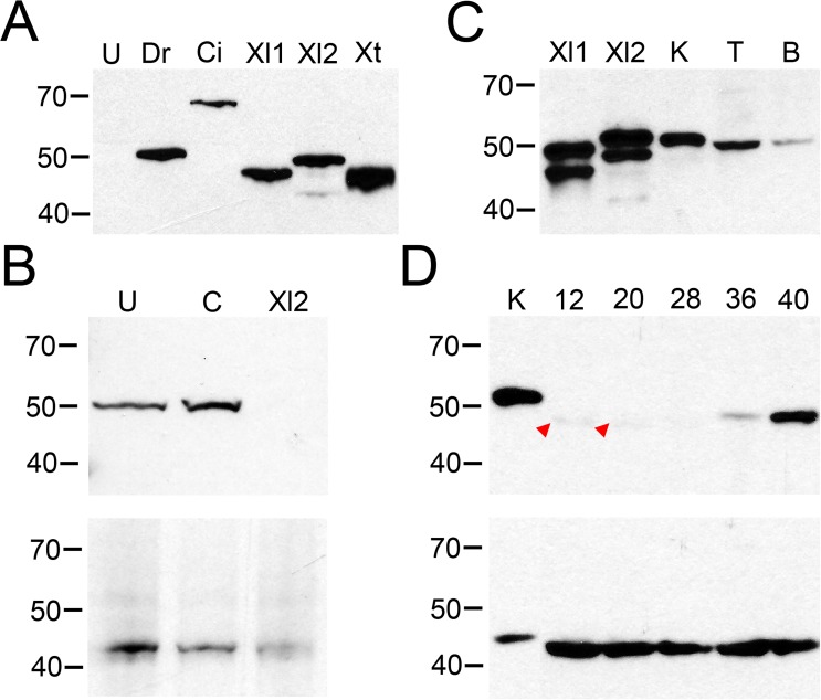
Fig 3. X. laevis tissues and embryos show VSP protein expression.
(A) Western blot validation of N432/21 anti-VSP in X. laevis oocytes injected with cRNA for Dr-VSP (Dr), FLAG-Ci-VSP (Ci), Xl-VSP1 (Xl1), Xl-VSP2 (Xl2), Xt-VSP (Xt), or left un-injected (U). All VSPs tested were recognized by the antibody. Un-injected oocytes (U) display no band. Dr-VSP, Xl-VSP1, Xl-VSP2 and Xt-VSP have a predicted MW of 58 kDa while FLAG-Ci-VSP has a predicted MW of 66 kDa. The slight difference in electrophoretic mobility between VSPs and their predicted MWs and the nature of the double band for Xl-VSP1 and Xl-VSP2 (as seen in panels A and C) has not been determined. These results show that anti-VSP N432/21 is specific for VSP and cross-reacts with VSPs from multiple species. (B) X. laevis zygotes were injected with either a control morpholino, an Xl-VSP2 morpholino or left un-injected. (top) Western blot analysis of equal amounts of lysates from embryos at stage 42 shows a band at the predicted MW for Xl-VSPs (58 kDa) for un-injected and control embryos and no band for the Xl2 morpholino-injected embryos. (bottom) Blots were stripped and re-probed with anti-actin for a loading control. (C) Western blot analysis of X. laevis tissues. Lysates from adult kidney (K, 3 μg), testis (T, 30 μg), and brain (B, 10 μg) were run against lysates from oocytes injected with RNAs for Xl-VSP1 (Xl1) and Xl-VSP2 (Xl2) and analyzed by Western blot with anti-VSP. A single band of approximately the correct MW (58 kDa) was observed in the tissue lysates, indicating the presence of Xl-VSP protein in all tissues tested. (D) Western blot analysis of X. laevis embryos. Lysates from NF stage 12–40 embryos (30 μg each) were run against a lysate from adult kidney (K, 5 μg). Blots were probed either with anti-VSP (top) or anti-actin (bottom) as a loading control (predicted MW 42 kDa). A weak band (potentially corresponding to Xl-VSP1) is present only at early embryonic stages 12–20 (red arrowheads), whereas a slightly slower-migrating band (potentially corresponding to Xl-VSP2) accumulates at later embryonic stages 36–40. The difference between the kidney and embryo lysate MWs for both VSP and actin may reflect a different degree of post-translational modification in embryos versus adult X. laevis. Lysates, gels and blots were repeated three times with either three different adults or three different embryonic cohorts. Shown are representative gels for each.

