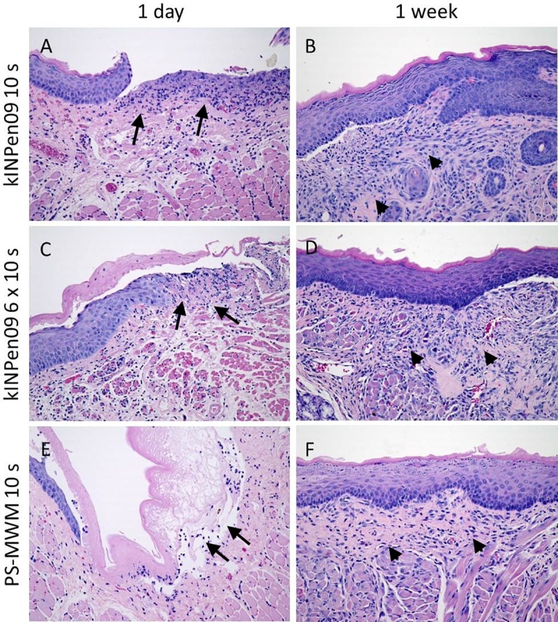
Fig 5. Histological examinations of plasma treated groups.

A, C, E: Cheek mucosa with ulceration and inflammation in the stroma (arrows) one day after the treatment with CAP (A kINPen09 10s, C kINPen09 6x10s, E PS-MWM 10s). B, D, F: Cheek mucosa one week after the CAP treatment (B kINPen09 10s, D kINPen09 6x10s, E PS-MWM 10s) with reepithelialization and remnants of granulation tissue (arrow heads). (A-F: HE, original magnification 200-fold).
