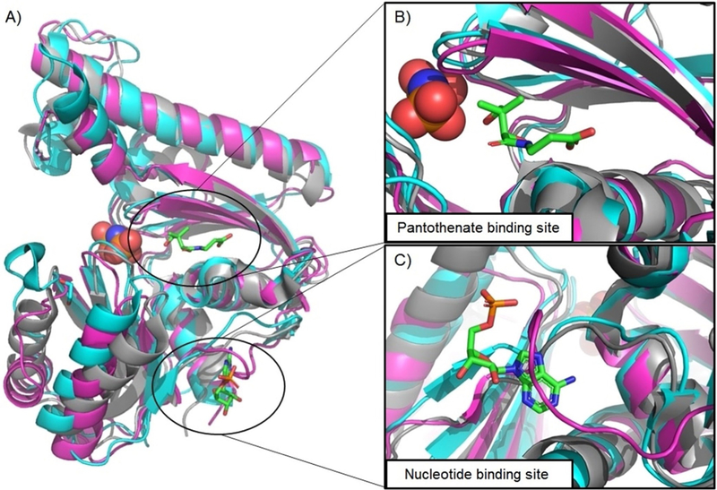
Figure 4.

A) Computed structural alignment of PanK crystal structures from Bacillus anthracis (magenta), Pseudomonas aeruginosa (grey), and Burkholderia cen- ocepacia (cyan). Pictured substrates (pantothenate, adenosine monophosphate, and imidodiphosphoric acid) are from the crystal structure of B. cenocepacia. B) Aligned structures of the pantothenate binding site, showing strong overlap. C) Aligned structures of the nucleotide binding site, showing a significant structural change in BaPanK relative to PaPanK and BcPanK, potentially explaining differential inhibition.
