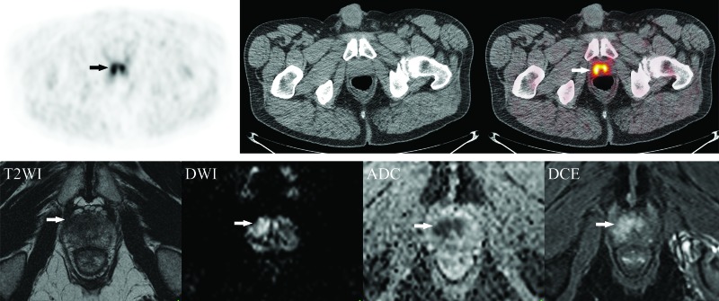
Fig. 2.
Example of local disease on 18F-fluorocholine positron emission tomography-computed tomography (18F-FCH PET/CT) with magnetic resonance imaging (MRI) correlation. (A) Axial 18F-FCH PET/CT images obtained in a 63-year-old man with prostate cancer (cT1c, Gleason score 8, prostate-specific antigen 12.6 ng/mL) showing bilateral prostate uptake (SUV 6.6) (arrows) without metastatic disease. (B) Corresponding axial MRI prostate images (from left to right: T2-weighted images [T2WI], diffusion-weighted imaging [DWI], apparent diffusion coefficient [ADC] map, and dynamic contrast-enhanced images [DCE]) demonstrating a non-circumscribed homogeneous moderately T2 hypointense lesion measuring 1.8 cm in maximal dimension located in the transition zone at the apex and mid-gland with mild extension to the right anterolateral peripheral zone (arrows). There is associated restricted diffusion on DWI/ADC and early focal enhancement on DCE (PI-RADS 5). The patient underwent radical prostatectomy with extended pelvic lymph node dissection (pT3a pN0), without evidence of biochemical recurrence after 10 months of followup. MRI images courtesy of Dr. F. Discepola, Jewish General Hospital, Montreal, QC, Canada.

