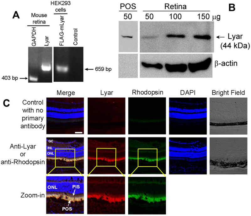
Fig. 3.

Lyar is expressed in the retina. (A) Lyar mRNA transcript in the retina was detected by RT-PCR. Housekeeping gene GAPDH was included as a positive control. HEK293 cells pre-transfected with Lyar-FLAG plasmid was used as a positive control. No template was used as a negative control. (B) Lyar expression in mouse retina or shed POSs was analyzed by Western blot with different amount of total retinal proteins. (C) Lyar expression in mouse retina was analyzed by immunohistochemistry. GC, ganglion cell; INL, inner nuclear layer; ONL, outer nuclear layer; PIS, photoreceptor inner segment.
