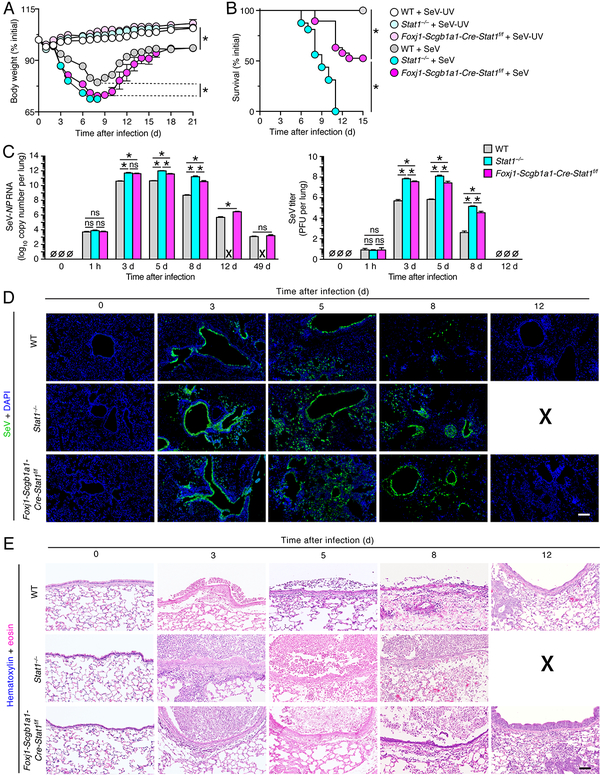
FIGURE 2.
STAT1 deficiency in epithelial cells increases severity of SeV infection. (A) Body weights for WT, Stat1−/−, and Foxj1-Scgb1a1-Cre-Stat1f/f mice at the indicated times after infection with SeV (1 × 10⁵ PFU given intranasally) or equivalent control SeV-UV. (B) Survival rates for conditions in (A). (C) Lung levels of SeV-NP RNA using PCR assay and corresponding levels of infectious SeV titers using plaque-forming assay for WT, Stat1−/−, and Foxj1-Scgb1a1-Cre-Stat1f/f mice at the indicated times after infection with SeV. (D) Immunostaining for SeV and counterstaining with DAPI in lung sections for conditions in (C). Scale bar, 400 μm. (E) Hematoxylin and eosin staining in lung sections for conditions in (C). Scale bar, 200 μm. For (A-E), values are representative of 3 separate experiments (n≥8 mice per condition in each experiment). For (A-B), * indicates p<0.05 by ANOVA except for survival rates by Kaplan-Meier analysis, and for (C-E), X signifies data that could not be obtained due to non-survival conditions.

