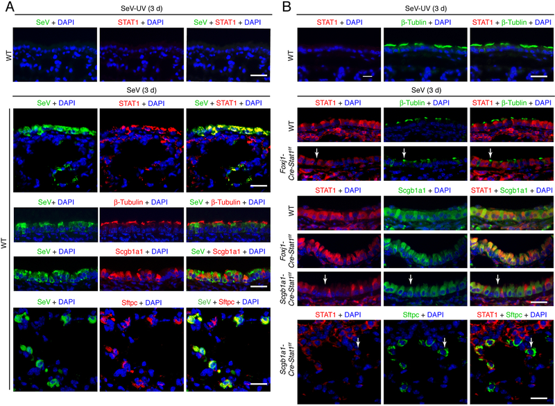
FIGURE 3.
SeV replication and STAT1 induction co-localizes to epithelial cells. (A) Immunostaining for SeV and STAT1, β-Tubulin IV, Scgb1a1, or Sftpc and counterstaining with DAPI in lung sections from WT mice at 3 d after infection with SeV. Scale bar, 100 μm. (B) Immunostaining for STAT1 and β-Tubulin IV, Scgb1a1, or Sftpc and counterstaining with DAPI in lung sections from Foxj1-Cre-Stat1f/f, Scgb1a1-Cre-Stat1f/f, and WT mice at 3 d after infection with SeV. Arrows indicate epithelial cells (green staining) without STAT1 (red staining) in the context of Stat1 gene deletion. Scale bar, 100 μm. For (A,B), values are representative of 3 separate experiments (n≥8 mice per condition in each experiment).

