FIGURE 6.
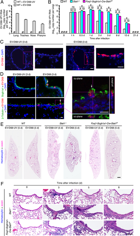
STAT1-deficiency in epithelial cells increases severity of EV-D68 infection. (A) Tissue levels of EV-D68-VP1 RNA in WT mice at 1 d after infection with EV-D68 (0.5 × 10⁶ ePFU given intranasally) or an equivalent amount of control EV-D68-UV. (B) Lung levels of EV-D68-VP1 RNA for WT, Stat1−/−, and Foxj1-Scgb1a1-Cre-Stat1f/f mice at the indicated times after infection with EV-D68. (C) Immunostaining for EV-D68 with DAPI counterstaining in lung sections from Stat1−/− mice at 3 d after infection with EV-D68 or EV-D68-UV. Scale bar, 100 μm. (D) Immunostaining for EV-D68 and Scgb1a1 or β-Tubulin IV and counterstaining with DAPI in lung sections for conditions in (C) imaged with conventional and confocal microscopy. Arrows indicate cells co-staining for EV-D68 and Scgb1a1 or β-Tubulin IV. Scale bars, 50 μm. (E) Hematoxylin and eosin staining in lung sections for conditions in (A). Scale bar, 500 μm. (F) Hematoxylin and eosin staining in airway sections for conditions in (A). Scale bar, 200 μm. For (A-E), data are representative of 3 separate experiments (n≥8 mice per condition in each experiment). For (A,C), * indicates p<0.05.

