FIGURE 9.
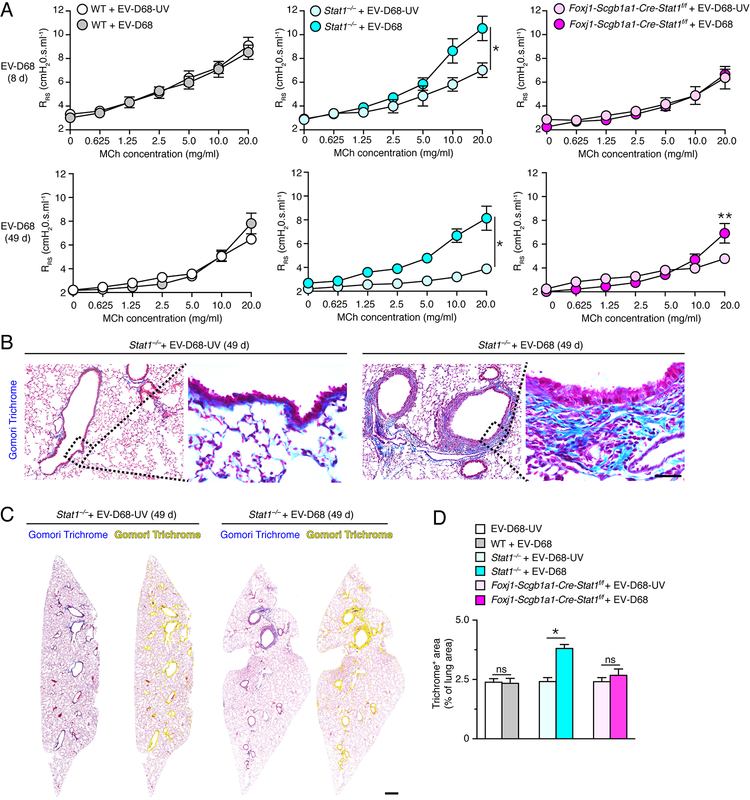
STAT1 deficiency in epithelial cells increases susceptibility to chronic lung disease marked by airway hyperreactivity and fibrosis after EV-D68 infection. (A) Levels of airway reactivity using response of respiratory system resistance (RRS) to inhaled methacholine (MCh) in WT, Stat1−/−, and Foxj1-Scgb1a1-Cre-Stat1f/f mice at 8 d and 49 d after infection with EV-D68 or EV-D68-UV. (B) Gomori trichrome staining of lung sections from Stat−/− mice at 49 d after infection with EV-D68 or EV-D68-UV. Scale bar, 200 μm. (C) Gomori trichome staining of whole lung sections for conditions in (B) with yellow colorization indicating computer-assigned collagen-containing light-blue+ staining areas. Scale bar, 500 μm. (D) Quantitation of trichome light-blue+ staining areas in lung sections for WT, Stat1−/−, and Foxj1-Scgb1a1-Cre-Stat1f/f mice at 49 d after infection with EV-D68 or EV-D68-UV. For (A,D), values are representative of 3 separate experiments (n=5–8 mice per condition in each experiment). For (A), * indicates p<0.05 by ANOVA for entire MCh concentration-response and ** indicates p<0.05 by post-hoc Bonferroni for single MCh concentration versus EV-D68-UV condition. For (D), * indicates p<0.05.

