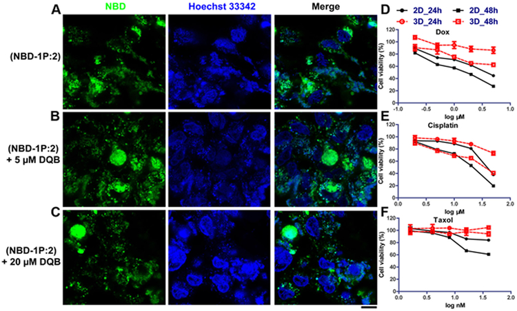
Figure 3. Extracellular iA of sPGP generates a biomimetic 3D model for drug screening.
Confocal images of Saos-2 cell lines treated with (A) NBD-1P plus 2 (300 μM) and (B) NBD-1P, 2 and DQB (5 μM) and (C) NBD-1P, 2 and DQB (20 μM) for 48 h. Scar bar is 10 μm. Cytotoxicity of three representative chemotherapy drugs (D) doxorubicin, (E) cisplatin, and (F) taxol on the 3D cell spheroids and the 2D cell culture.

