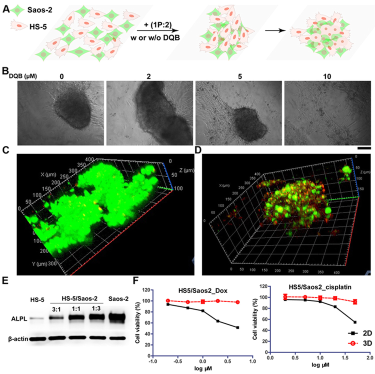
Figure 4. iA of sPGP induces formation of spheroids of heterotypic cells.
(A) The illustration of the formation of 3D cell spheroids made of HS-5 and Saos-2 cells from a 2D cell sheet upon the addition of 1P:2. (B) Microscope images of the co-culture of HS-5 and Saos-2 (ratio is 1:1) cells at the density of 3×10⁴ treated with mixture of 1P:2 (300 μM) with different concentration of DQB for 48 h. Scar bar is 150 μm. (C) CLSM images of live-dead assay of the co-culture of HS-5 and Saos-2 cells treated with mixture of 1P:2 (300 μM) for 48 h. (D) CLSM images of the co-culture of HS-5 and Saos-2 (ratio is 1:1) cells at the density of 3×10⁴ treated with mixture of 1P:2 (300 μM) for 48 h. HS-5 cell lines were treated with Hoechst 33342 (red) for 10 minutes prior to co-cultur with the Saos-2 cell that were treated with membrane probe (green) for 1 h. (E) Expression levels of ALPL on HS-5, Saos-2, and the mixture of HS-5 and Saos-2 at different ratios. The total cell numbers in each group are same. (F) Cytotoxicity of two representative chemotherapy drugs (doxorubicin and cisplatin) on co-cultured 3D spheroids and 2D cell culture for 24 h.

