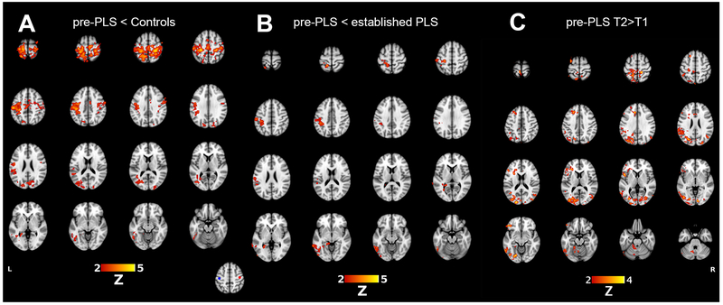
Figure 1.
Motor resting-state networks, showing differences in connectivity of the right motor cortex seed to the rest of the brain. (A) Red indicates brain regions with reduced connectivity in pre-PLS patients compared to heathy controls. (B) Red indicates brain regions in which established PLS patients have greater connectivity than in pre-PLS patients. (C) For eight pre-PLS patients with follow-up scans, regions in red had greater connectivity with the right motor cortex seed at the second timepoint (T2) compared to the first study (T1). Inset figure: 10-mm VOIs for generating the sensorimotor resting-state network were obtained from the voxel of peak activation in the left (MNI: −42, −20, 52; blue circle) and right (42, −16, 52; red circle) motor cortex from the right and left hand tapping task fMRI.

