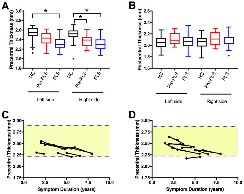
Figure 2.
Thinning of the motor cortex in PLS patients.
(A) The mean thickness of the precentral gyrus was reduced in both hemispheres in established PLS patients (blue) compared to healthy controls (HC, black). Slight thinning of the right precentral cortex was seen in pre-PLS patients (red) at baseline. Asterisks indicate Bonferroni-corrected p < 0.05. Age, gender, and education were included as covariates. (B) In contrast, the postcentral gyrus had no significant thinning in PLS and Pre-PLS patients. (C, D) Longitudinal measures of cortical thickness of the precentral gyrus of 8 pre-PLS patients who had repeated scans. Cortical thickness is plotted against years since symptom onset. (C) Left precentral gyrus and (D) right precentral gyrus. Shaded area indicates the range of thickness for the healthy controls (mean ± 2 SDs).

