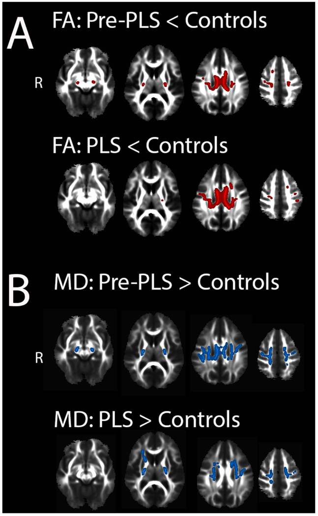
Figure 3.
White matter changes in patients compared to controls.
Regions of white matter with (A) reduced fractional anisotropy (red) in pre-PLS patients (top row) and established PLS patients (bottom row) compared to healthy controls. Regions of white matter with (B) increased mean diffusivity (blue) in pre-PLS patients (top row) and established PLS patients (bottom row) compared to healthy controls. There was no significant difference between pre-PLS and PLS patients (Tract-based spatial statistics, p < 0.05, FWE corrected.)

