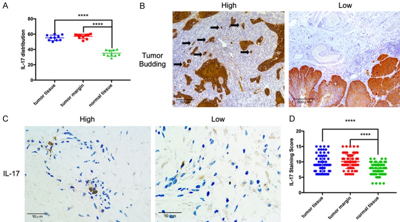
Figure 1.

IL-17 and pan-cytokeratin expression in oral squamous cell carcinoma (OSCC) tissues. A. IL-17 levels in tumor tissues, tumor margins, and normal tissues evaluated by ELISA. B. Left: Immunohistochemistry staining of pan-cytokeratin in high-grade tumor budding; black arrows indicate tumor budding. Right: Low-grade tumor budding. C. Immunohistochemistry staining of IL-17 showing high and low expression levels in the tumor invasion front. D. The IL-17 expression was significantly elevated in tumor tissues and tumor margins compared with that in normal tissues. ****P < 0.001.
