
Figure 5.
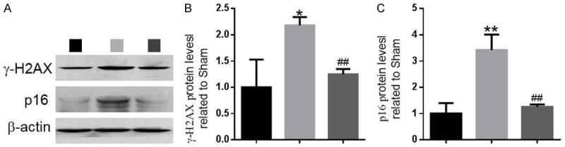
The effect of PQQ on DNA damage and cellular senescence in articular cartilage of ACLT-induced OA mice. (A) Representative western blots of articular cartilage extracts analysed for expression of γ-H2AX and p16INK4a. β-actin was used as loading control for western blots in the sham, ACLT and ACLT+PQQ mice. (B) γ-H2AX and (C) p16INK4a protein levels relative to β-actin protein levels were assessed by densitometric analysis and expressed relative to levels in sham mice. Data are presented as the mean ± SEM of determinations; each data-point is the mean of five specimens. *P < 0.05, **P < 0.01 versus sham mice. ##P < 0.01 versus PQQ-untreated ACLT mice.