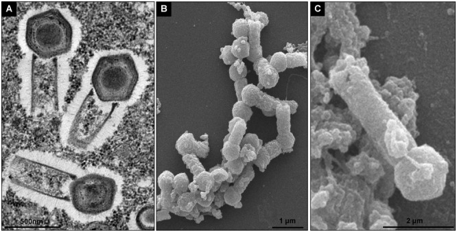
FIGURE 1.

Tupanvirus soda lake particle. (A) Mature particle of Tupanvirus soda lake (TPVsl) in V. vermiformis under transmission electron microscopy (TEM). (B) Mature particle of TPVsl in V. vermiformis under scanning microscopy. Is it possible note the peculiar TPVsl morphology, with the tail attached to a Mimivirus-like capsid. (C) “Supertupan” in V. vermiformis under scanning electron microscopy.
