FIGURE 4.

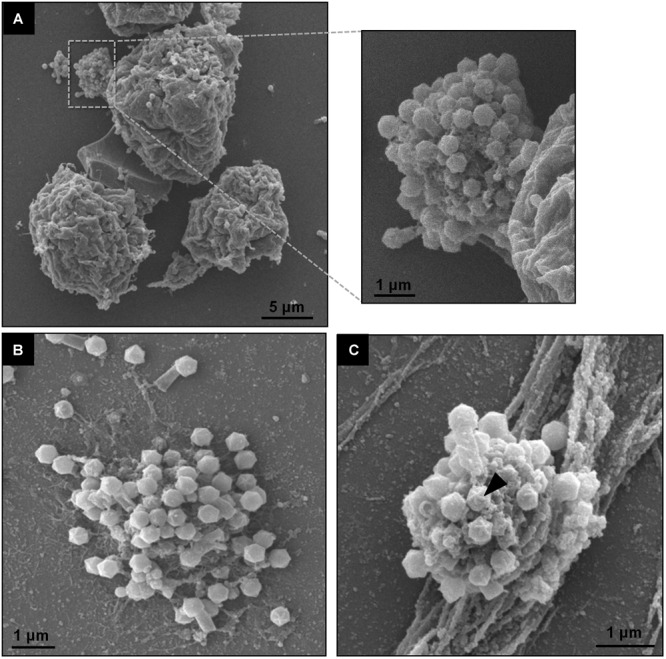
TPVsl viral factory by scanning microscopy. (A) Details of a mature VF involved in viral morphogenesis, including tail attachment. (B,C) Isolated VFs releasing viral particles. The stargate in a capsid is indicated by the black arrow.

TPVsl viral factory by scanning microscopy. (A) Details of a mature VF involved in viral morphogenesis, including tail attachment. (B,C) Isolated VFs releasing viral particles. The stargate in a capsid is indicated by the black arrow.