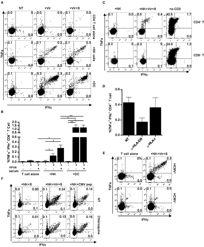
Figure 4.
CD4+ T cell activation in response to HCMV antigen presentation by NK or moDC. NK cells or moDCs previously loaded with HCMV-antibody immune complexes were cultured overnight with autologous CD4+ T cells. TNFα and IFNγ production was analyzed by flow cytometry. (A) TNFα and IFNγ production by CD4+ T cells in the indicated conditions. Data from a representative donor out of five tested. (B) Mean frequency of IFNγ+ and TNFα+ CD4+ T cells upon activation with different APCs (mean ± SEM, n = 5) (*p < 0.05, **p < 0.01). (C–E) Autologous PBMC were used as effectors in co-culture experiments with NK cells pre-incubated with HCMV-antibody immune complexes. CD4+ and CD8+ T cell activation was analyzed by flow cytometry. An agonist anti-CD3 antibody was used as a positive control. (C) Dot plots display intracellular TNFα and IFNγ in CD4+ and CD8+ T cells in the indicated conditions. Data from a representative donor out of four analyzed. (D) Frequency of TNFα+ and IFNγ+ CD4+ T cells in response to HCMV-loaded NK cells in the presence of blocking antibodies specific for HLA-DR and HLA class I molecules (mean ± SEM, n = 3). (E) TNFα and IFNγ intracellular staining of CD4+ T cells in co-cultures including antigen-presenting NK cells and autologous PBMC from HCMV seropositive and seronegative individuals. (F) Frequency of TNFα+ and IFNγ+ CD4+ T cells in response to HCMV-loaded NK cells. NK cells were loaded in the presence or absence of chloroquine (50 μM). Dot plots of one out of two donors tested.

