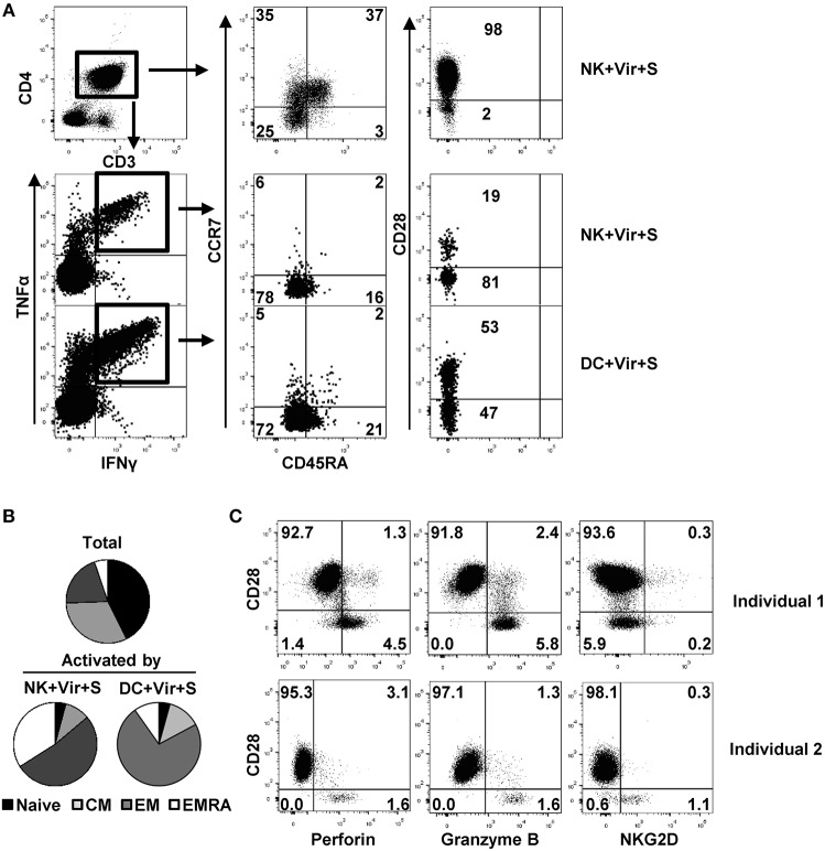
Figure 5.
Differentiation and functional profile of HCMV-specific CD4+ T cells activated by antigen-presenting NK cells. NK cells or moDC previously loaded with HCMV in the presence of specific antibodies were cultured with autologous CD4+ T cells and the production of TNFα and IFNγ in combination with CD45RA, CCR7, and CD28 differentiation markers was analyzed by multiparametric flow cytometry. (A) Dot plots showing CD45RA, CCR7, and CD28 expression in total and activated (IFNγ+ TNFα+) CD4+ T cells from a representative donor out of four in the indicated conditions. (B) Pie chart showing the distribution of CD4+ T cell subpopulations based on CCR7 and CD45RA at baseline and of those T cells activated by antigen-presenting NK cells and DC (n = 4). (C) Perforin, granzyme B, and NKG2D expression in CD28+ and CD28– CD4+ T cells from two representative HCMV+ individuals out of three analyzed.

