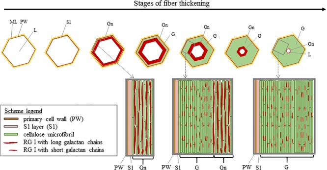
FIGURE 6.

Scheme illustrating the cell wall thickening in relation to the remodeling of the Gn-layer. Cellulose microfibrils are originally separated by long galactan chains present in nascent RG I [5]. The long galactan chains are then trimmed off which leads to the G-layer with compact cellulose microfibrils. Scheme inspired from Goudenhooft et al. (2018).
