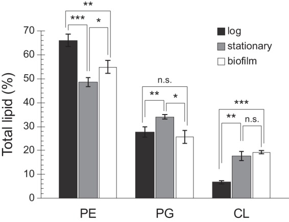
FIG 2.

CL is elevated in cells from early-stage biofilms compared with cells in log phase. Total membrane lipids were isolated from MG1655 cells grown in suspension to an absorbance (λ, 600 nm) of 0.4 to 0.6 (log) for 24 h (stationary) or in 96-well plates without shaking for 24 h (biofilm). After separation by TLC, phospholipids were visualized by fluorescence imaging following treatment with cupric sulfate. ImageJ was used to quantitate lipid spots. Error bars indicate standard deviation of 3 biological replicates; ***, P < 0.001; **, P < 0.01; *, P < 0.05; n.s., not significant; Student’s t test.
