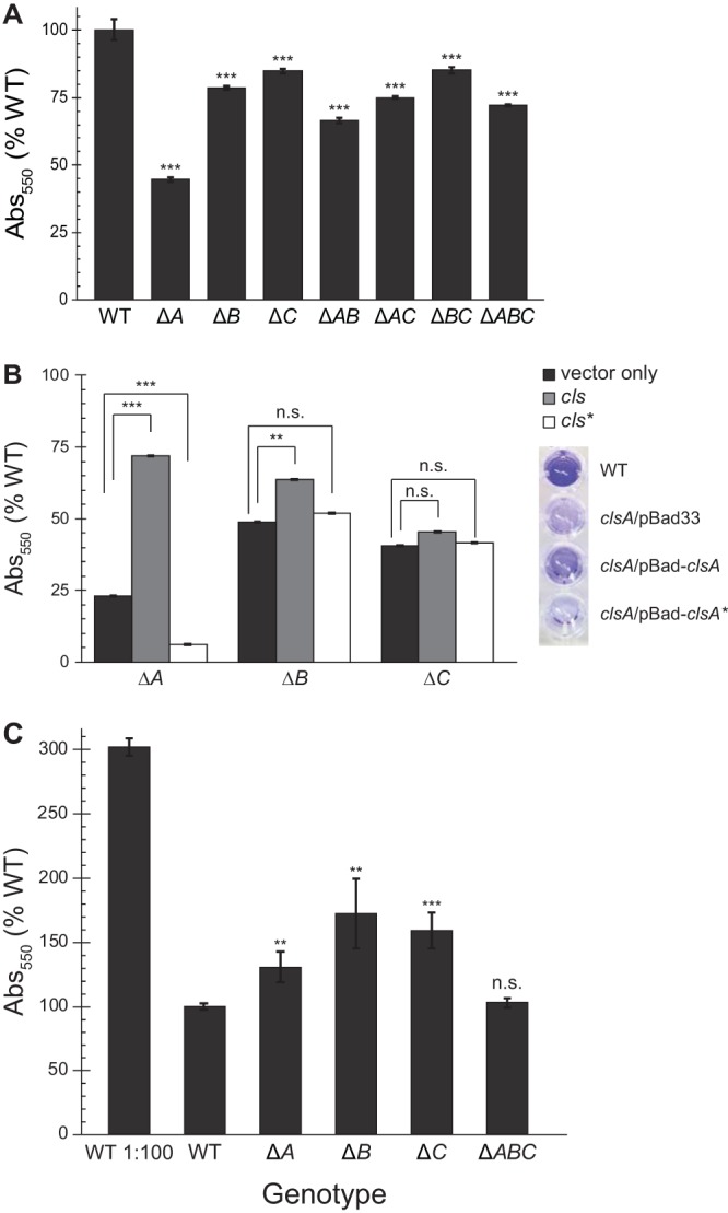
FIG 3.

Disruption of cardiolipin synthesis reduces biofilm formation. E. coli cells were grown in microtiter plates for 24 h at 30°C without shaking. Adherent cells were stained with crystal violet (CV), and CV absorbance was measured (λ, 550 nm). Error bars indicate standard error; ***, P < 0.001; **, P < 0.01; *, P < 0.05; n.s., not significant; Student’s t test compared with WT. (A) CV labeling of mutant E. coli cells with noted genotypes. (B) Δcls strains were complemented with pBad33 (vector), full-length cls, or a catalytically inactive mutant (cls*; clsA H224F H404F, clsB H113A, or clsC H130A). Cells were grown with 0.2% arabinose to induce protein expression; CV absorbance values of cells grown with arabinose were normalized to CV absorbance of uninduced cells. Error bars indicate standard error; ***, P < 0.001; **, P < 0.01; n.s., not significant; Student’s t test compared with uninduced control. Right, image of representative crystal violet staining of cells grown in M9 with 0.2% arabinose. From top, wild-type (MG1655), ΔclsA/pBad33, ΔclsA/pBad-clsA, and ΔclsA/pBad-clsA* strains. (C) CV staining of biofilms started using saturated overnight cultures; CV labeling of biofilm started with subcultured WT is shown for reference. Error bars indicate standard error; ***, P < 0.001; **, P < 0.01; n.s., not significant; Student’s t test compared with WT.
