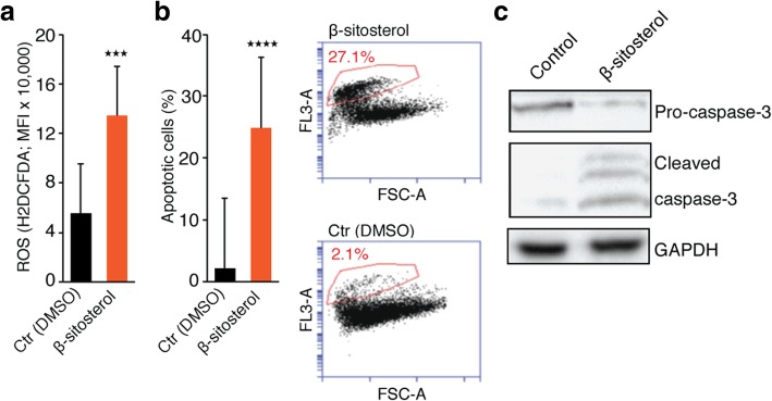
Fig. 6.
β-sitosterol increases ROS production and apoptosis. a ROS content (CM-H2DCFDA probe; mean fluorescence intensity (MFI); n = 2 with triplicates). b Flow cytometric apoptosis assay (n = 3) showing a strong induction of apoptosis following ß-sitosterol treatment. c Western blot of pro-caspase-3, cleaved caspase-3 and GAPDH in H1_DL2 cells exposed to DMSO (0.05%) or β-sitosterol (50 μM) for 2, 24 or 24 h, respectively. Student’s t-test: *** P < 0.001, **** P < 0.0001. Values are given as the mean ± s.e.m

