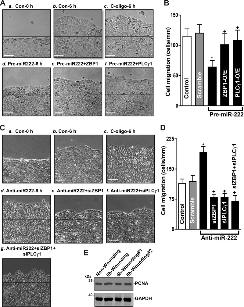
Fig. 6.
microRNA-222 (miR-222)-regulated expression of zipcode binding protein-1 (ZBP1) and phospholipase C-γ1 (PLCγ1) modulates rapid epithelial restitution after wounding. A: images of cell migration: a, 0 h after wounding in control cells (Con-0 h); b, 6 h after wounding in control cells (Con-6 h); c, 6 h after wounding in cells transfected with scramble oligo (C-oligo-6 h); d, 6 h after wounding in cells transfected with pre-miR-222 alone for 48 h (Pre-miR-222–6 h); e, 6 h after wounding in cells cotransfected with pre-miR-222 and the expression vector encoding ZBP1 (Pre-miR222 + ZBP1); f, 6 h after wounding in cells cotransfected with pre-miR-222 and the expression vector encoding PLCγ1 (Pre-miR222+ PLCγ1). Scale bar, 100 μm; magnification, ×100. B: summarized data showing rates of cell migration 6 h after wounding in cells described in A. Values are the means ± SE of data from 6 dishes and repeated 4 times independently (n = 4). *,+P < 0.05, compared with cells transfected with scramble and cells transfected with pre-miR-222, respectively as analyzed by one-way ANOVA followed by Duncan’s test. C: images of cell migration: a: 0 h after wounding in control cells; b: 6 h after wounding in control cells; c, 6 h after wounding in cells transfected with scramble oligo; d: 6 h after wounding in cells transfected with anti-miR-222 alone for 48 h; e: 6 h after wounding in cells cotransfected with anti-miR-222 and siZBP1 (Anti-miR222 + siZBP1); f: 6 h after wounding in cells cotransfected with anti-miR-222 and siPLCγ1 (Anti-miR222 + siPLCγ1); and g: 6 h after wounding in cells cotransfected with anti-miR-222, siZBP1 and siPLCγ1 (anti-miR222 + siZBP1 + siPLCγ1). Scale bar, 100 μm; magnification, ×100. D: summarized data showing rates of cell migration in cells described in C. Values are the means ± SE of data from 6 dishes and repeated four times independently (n = 4). *,+P < 0.05, compared with cells transfected with scramble and cells transfected with Anti-miR-222, respectively as analyzed by one-way ANOVA followed by Duncan’s test. E: immunoblot of PCNA protein in nonwounding and 0 h and 6 h after wounding (#1 and #2). Whole cell lysates were harvested and prepared for Western blotting; equal loading was monitored by assessing GAPDH levels.

