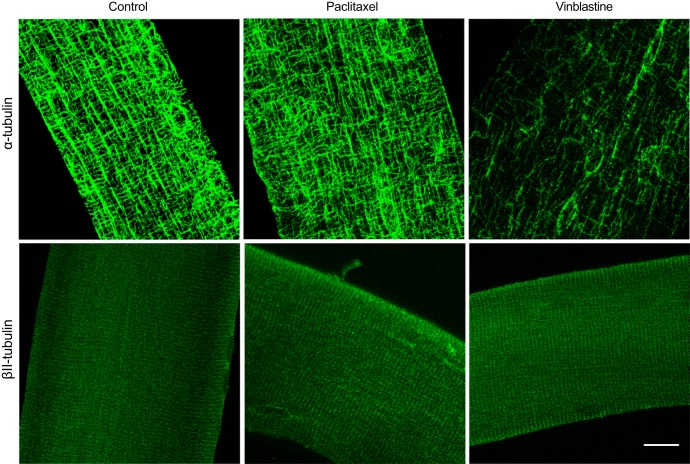
Fig. 2.
α-Tubulin but not βII-tubulin organization is altered following microtubule-targeted chemotherapy. Confocal microscopy representative images of single extensor digitorum longus fibers stained with antibodies recognizing α-tubulin and βII-tubulin following either control (DMSO), paclitaxel, or vinblastine treatment. Scale bar, 16 μm.

