. 2019 Feb 27;10(14):3976–3986. doi: 10.1039/c8sc05771k

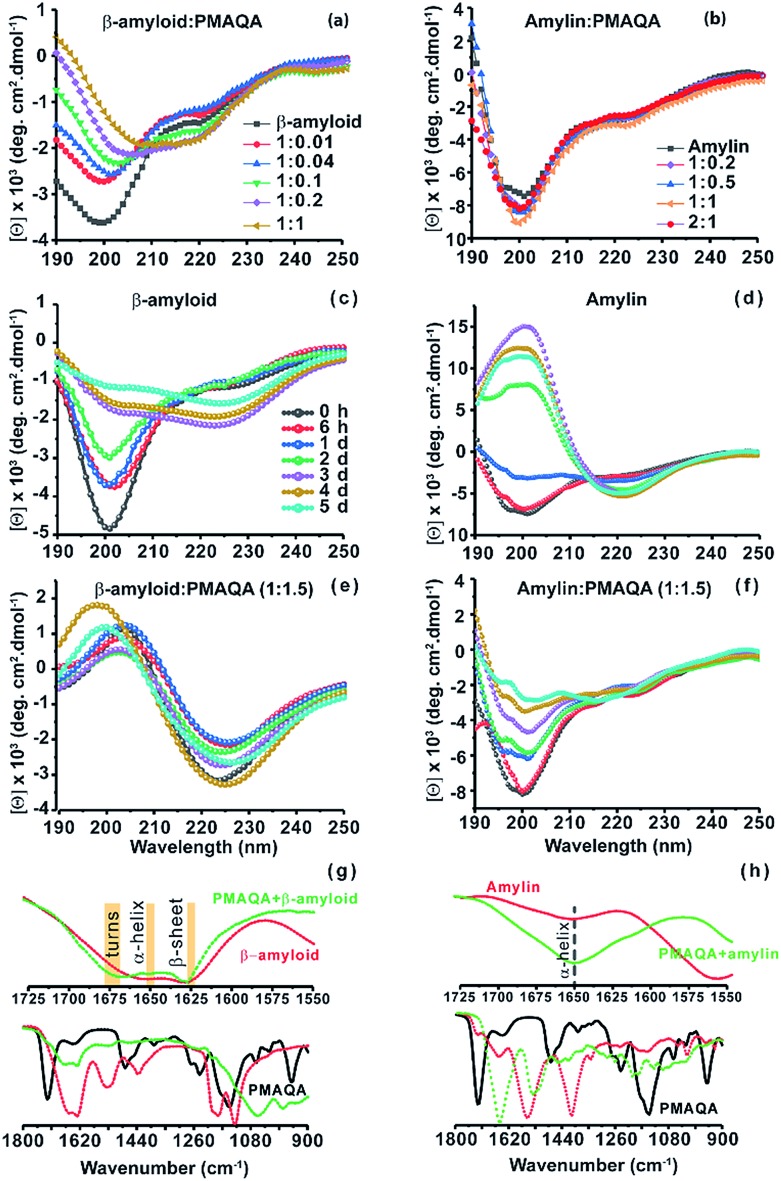
Fig. 2. Conformational analysis of β-amyloid-1-40 and amylin with and without PMAQA. Far-UV CD measurements showing the structural changes in 25 μM of β-amyloid-1-40 in 10 mM sodium phosphate, pH 7.4, (a) and in 25 μM of amylin in 30 mM sodium acetate, pH 5.5, (b) in the presence of PMAQA incubated for ∼10 minutes at room temperature at the indicated peptide : PMAQA molar ratios (a and b). Time-lapsed structural changes in 25 μM β-amyloid-1-40 (c and e) and amylin (d and f) in the absence (c and d) and presence (e and f) of 1.5 molar equivalent PMAQA; time intervals used for results presented in c–f are indicated in (c). FT-IR spectra of β-amyloid-1-40 (g) and amylin (h) in the presence (green) and absence (red) of PMAQA obtained after 6 hours; peptide concentration and buffer conditions were as described for the samples used in CD measurements. Changes in the secondary structure of β-amyloid-1-40 are highlighted in yellow in the fingerprint regions (1550–1725 cm–1) (g, top).

Fig. 2