. 2019 Feb 27;10(14):3976–3986. doi: 10.1039/c8sc05771k

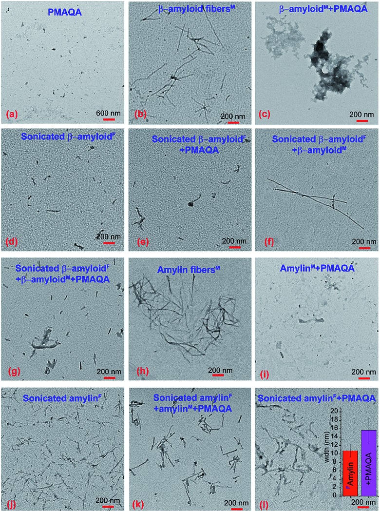
Fig. 7. TEM images of β-amyloid-1-40 and amylin interacting with PMAQA. TEM images of 5 μM PMAQA (a) and 5 μM β-amyloid-1-40 in the absence (b) or presence (c) of an equivalent concentration of PMAQA incubated for 24 hours at room temperature under continuous agitation. TEM images of sonicated β-amyloid-1-40 fibers (d) prepared from matured fibers (see the Experimental section) in the presence of 5 μM PMAQA (e); 5 μM β-amyloid-1-40 monomers (f); and 5 μM PMAQA and 5 μM β-amyloid-1-40 monomers (g) incubated for 24 hours at room temperature under continuous agitation. TEM images of 5 μM amylin in the absence (h) or presence (i) of 5 μM PMAQA incubated for 24 hours at room temperature under continuous agitation. TEM images of sonicated amylin fibers (j) prepared from matured fibers (see the Experimental section) in the presence of 5 μM PMAQA and 5 μM amylin monomers (k) and 5 μM PMAQA (l) incubated for 24 hours at room temperature under continuous agitation. The superscripts “M” and “F” denote the initial species of the peptide as monomers and fibers, respectively, used for the sample preparation. The inset in (l) shows the average width of amylin fibers that were calculated from 10 distinct short amylin fibers selected randomly using the ImageJ plugin from (j) and (l).

Fig. 7