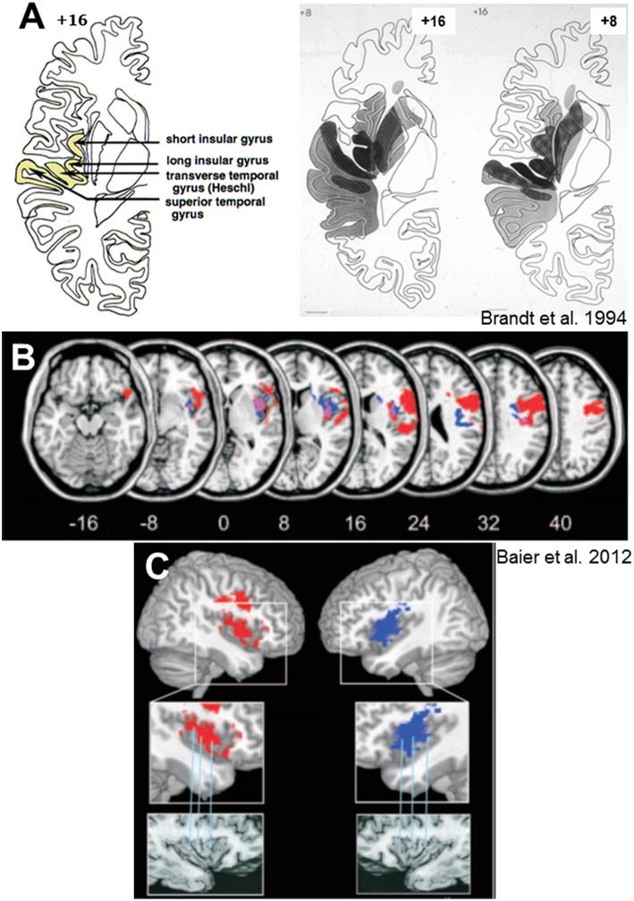
Figure 5.
Lesion sites of hemispheric infarctions that cause tilts of subjective visual vertical. (A) Collective presentation of infarcted areas taken from MRI scans and projected onto sections of the atlas of Duvernoy (81) in 7 patients with clearly demarcated infarctions of the middle cerebral artery which caused significant contralateral SVV tilts. Overlapping areas of infarctions (7 of 7 in black) are centered at the posterior part of the insula, involving the short and long insular gyri, the transverse temporal gyrus, and the superior temporal gyrus [from Brandt et al. (55)]. (B) Statistical voxelwise lesion-behavior mapping (VLBM) analysis comparing 32 patients with acute right-sided infarctions (RBD) and 22 patients with acute left-sided infarctions (LBD) with respect to absolute tilt of subjective visual vertical (t-test statistic). Presented are all voxels that survived a correction for multiple comparisons using a 1% false discover rate cutoff threshold. Overlay of the statistical map from LBD patients (blue color), flipped to the right hemisphere, and the statistical map of the RBD patients (red color). Overlapping regions are shown in violet. From Baier et al. (79) (C) Illustration of the affected parts of the insula using the atlas of Duvernoy (81). Right insular lesions in red, left insular lesions in blue. Affected are the circular insular sulcus, central insular sulcus, short insular gyrus, and long insular gyrus [From Baier et al. (79)].

