Figure 3.
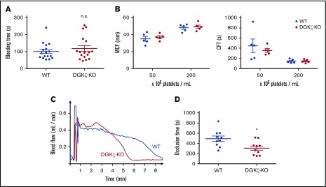
DGKζ-KO mice exhibit normal hemostasis but faster time to platelet plug formation after arterial injury. (A) Bleeding assay was performed by tail tip amputation followed by immersion in phosphate-buffered saline at 37°C. Symbols represent times to cessation of bleeding for individual animals, and error bars represent the mean ± SEM (n = 10-12). (B) ROTEM was performed using whole blood collected from WT and DGKζ-KO mice and adjusted to the indicated platelet count. Results are shown as maximal clot firmness (MCF; n = 5; left) and clot formation time (CFT; n = 5; right). Symbols represent MCF and CFT for individual animals, and bars represent the mean ± SEM. (C-D) The right carotid artery was injured by applying 10% FeCl3 for 3 minutes. Blood flow was monitored with a Doppler flow probe until 3 minutes after full occlusion. (C) Representative flow traces for WT and DGKζ-KO mice. (D) Symbols represent times to occlusion in individual animals, and bars represent the mean ± SEM (n = 10). Statistical analysis was performed by the unpaired Student t test. *P < .05 of DGKζ-KO as compared with WT.

