
Figure 2.
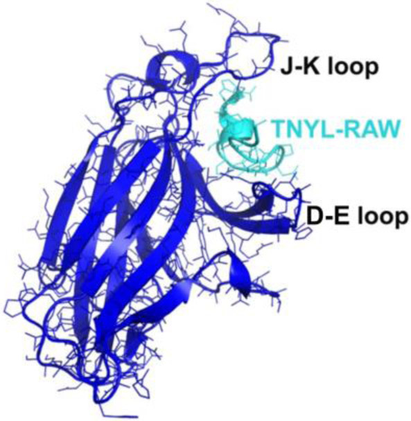
Ligand-Binding Domain (LBD) of the EphB4 receptor (in blue) in complex with an antagonistic peptide, TNYL-RAW (in cyan) (Chrencik et al., 2006). LBD has a jellyroll folding topology with 13 antiparallel B-sheets connected by several loops of varying lengths. Binding of TNYL-RAW makes the fairly flexible D-E and J-K loops more structured and they can be visualized in the electron density map.