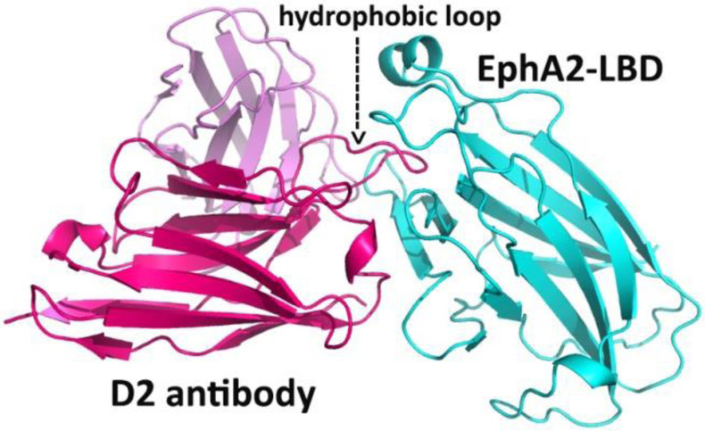
Figure 3.

Crystal structure of an anti-EphA2 scFv Ab in complex with the ligand-binding domain of EphA2. The scFv variable light chain is pink, the variable heavy chain is red, and the EphA2 receptor is cyan. A long loop of the D2 heavy chain penetrates a hydrophobic surface cavity of the EphA2 in a way very similar to Ephrin binding to Eph.
