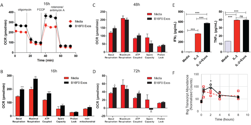
Figure 8: Exosomes increased aspects of mitochondrial respiration and IFN-γ production in CTLL2 cells.
(A) Oxygen consumption rate (OCR) in CTLL2 cells treated with culture media (red circles) or media containing B16F0 exosomes (black circles) was measured after 16 hour culture while the indicated chemical inhibitors of the respiratory chain were sequentially added. As described in the methods, metrics associated with mitochondrial respiration were inferred from the trace of the OCR after 16 hours (B), 48 hours (C) and 72 hours (D). Significance associated with the difference in basal respiration, maximal respiration, ATP-coupled respiration, non-mitochondrial respiration, space capacity and proton leak in exosome treated (black bars) compared to untreated cells (red bars) were assessed. (E) IFN-γ and TNF-α were assayed in CTLL2 conditioned media by cytometric bead array following the indicated treatments. (F) RNA-seq results for IFN-γ mRNA are shown for comparison. Results representative of two independent experiments that each contained at least four biological replicates, where ***, **, and * correspond to p-values calculated using an unpaired t-test of < 0.001, < 0.01, and < 0.05, respectively.

