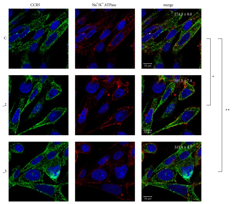
Figure 2.
Localization of CCR5 in PC12 cell lines. PC12 cells differentiated for 48 h with db-cAMP were fixed and immunostained with antibodies against CCR5 receptor (green) and Na+/K+ ATPase (plasma membrane marker, red). Nuclei were stained with Hoechst 33342 (blue). Representative confocal images are presented. Values shown in merged images represent average fluorescence intensity ± SEM (n = 5) of pixels positive in green channel (CCR5) that colocalize with red channel positive pixels (Na+/K+ ATPase). ∗ P< 0.05, ∗∗ P < 0.01. Scale bars: 10 μm. C: control line, _2: PMCA2-reduced line, _3: PMCA3-reduced line.

