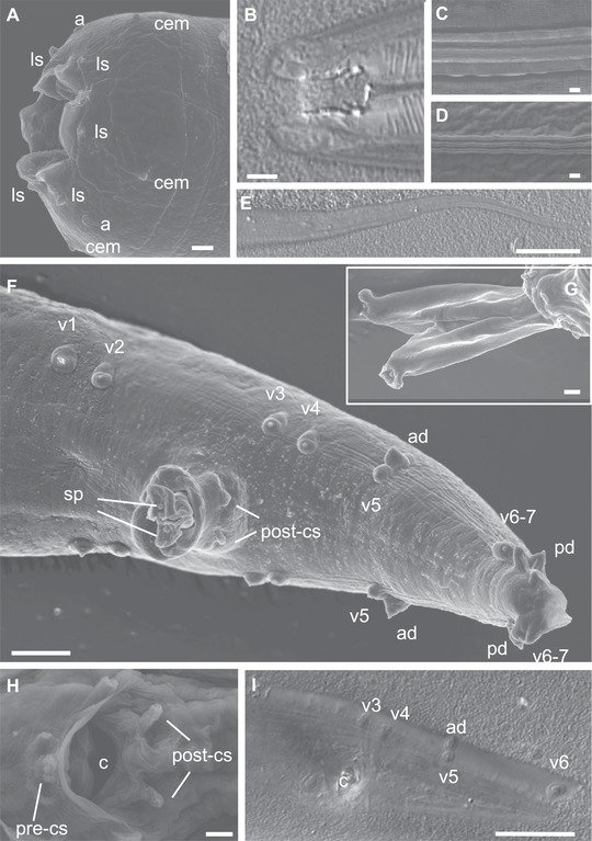
Figure 2.

Morphology of Caenorhabditis parvicauda sp. n. by scanning electron microscopy (SEM) and Nomarski optics (DIC). (A and B) Mouth of an adult male (A, SEM; B, DIC). (C) Cuticular lateral ridges of a dauer juvenile (SEM). (D) Cuticular lateral ridges of an adult male (SEM). (E) Female tail (DIC). (F) Male tail, ventro‐lateral view (SEM). (G) Genital opening with extruded spicules. (H) Male genital opening. (I) Male tail in ventral view (DIC). Anterior is to the left in (A, B, E–I). The animals are from strain JU2070. a, amphid; ad, anterior dorsal papilla; c, cloaca; cem, male cephalic sensillum (absent in females); ls, labial sensillum; pd, posterior dorsal papilla; pre/post‐cs, pre/post cloacal sensillum; v1, etc.: ventral papilla 1, etc.; sp, spicule. Scale bars: 1 μm, except in (E, F, and I): 5 μm. See also Figure S6.
