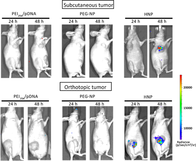
Figure 7.
HNPs maintains its gene delivery capability in mouse mammary xenograft tumor tissues while PEG-NP and PEI25k lost that. The mouse mammary tumors were produced as described in the Experimental Section. A single dose of HNPs loaded with 25 μg of Luc2 expressing pDNA was introduced into tumor tissues through an intratumoral injection. Twenty-four and forty-eight hours later, the animal was given D-luciferin by intraperitoneal injection, and the activity of tumor tissue-expressed Luc2 was determined by measuring the intensity of chemiluminescence signals using an IVIS live animal bioluminescence imaging system as described in the Experimental Section. The photo is a representative image showing strong chemiluminescence signals in mouse mammary tumor tissue (n = 3).

