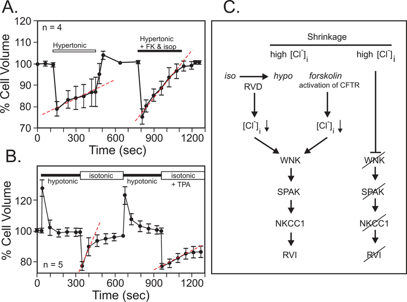
Figure 11. Intracellular Cl− and NKCC1 account for RVI in cells.

A, Behavior of simian eccrine clear cells exposed to hypertonic solution. Minimal RVI is observed unless the cells were treated with forskolin (FK) and isoproterenol (isop). B, Post-RVD RVI is strong in these cells but inhibited by phorbol ester (TPA). Data were redrawn and modified from (Toyomoto, Knutsen, Soos & Sato, 1997). C, Both RVD and forskolin through activation of Cl− channels cause a loss of intracellular Cl−. Through the activation of the protein kinases WNK and SPAK, this leads to an active NKCC1, which helps the cells recover their volume. Under non-stimulated conditions, the high intracellular Cl− prevents activation of Na-K-2Cl cotransport and the cells remain shrunken.
